Wednesday, November 14, 2012
Thursday, April 19, 2012
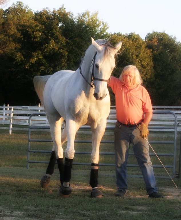
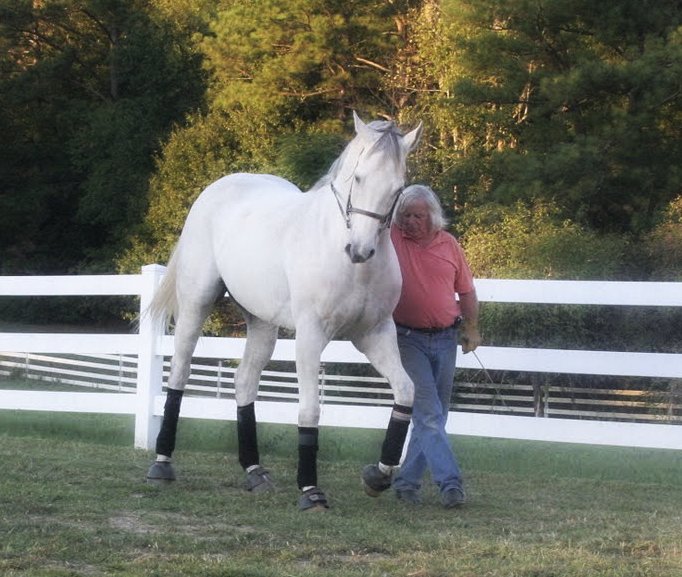
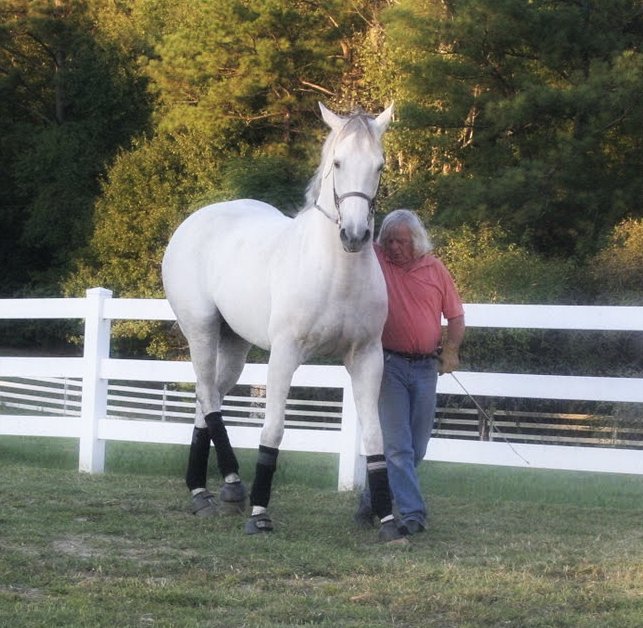
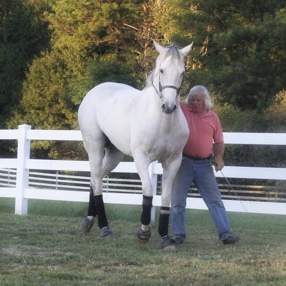
Friday, April 06, 2012
Saturday, March 24, 2012

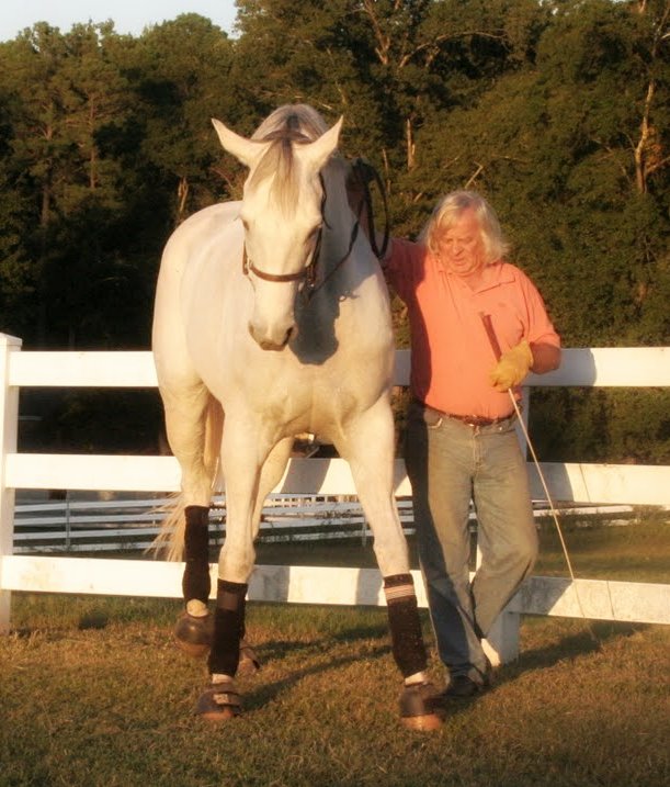
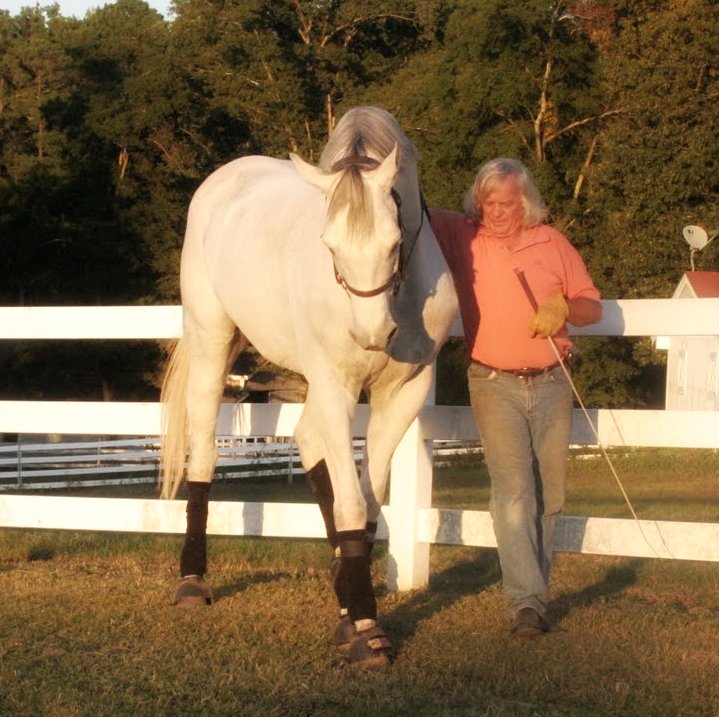
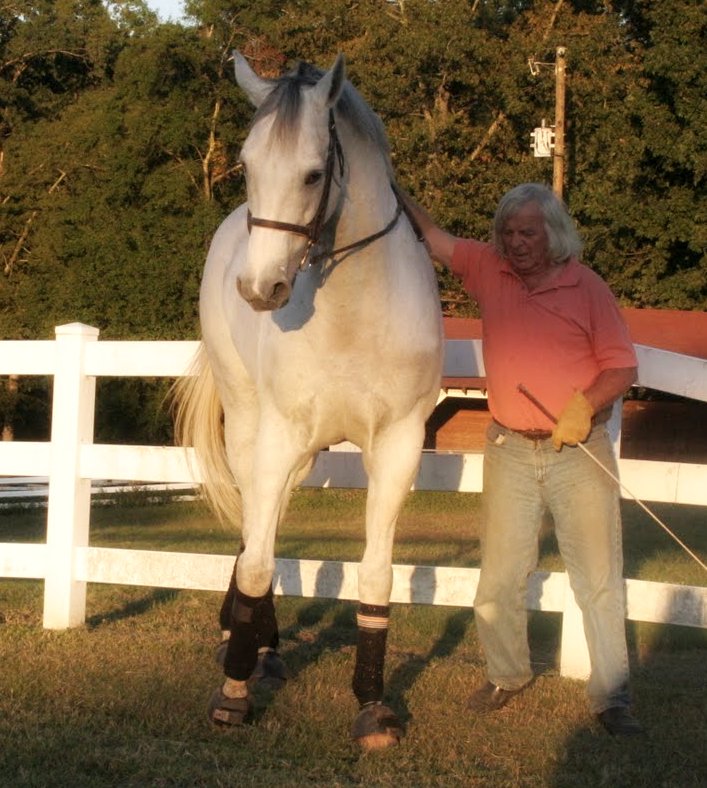
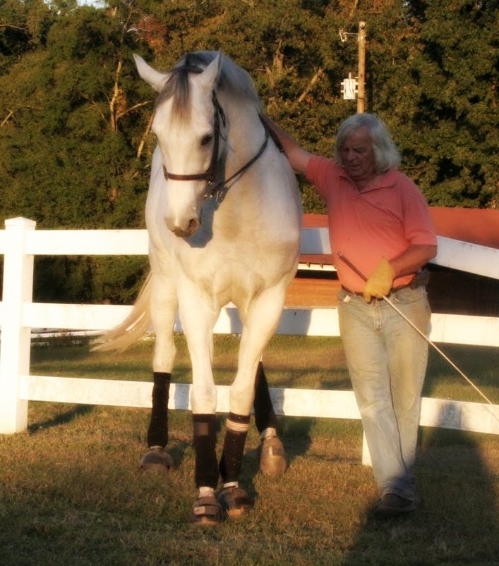
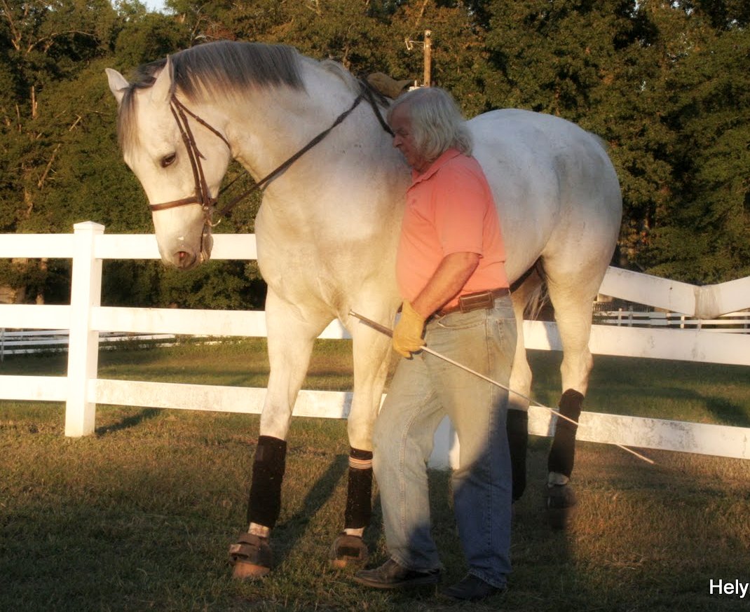
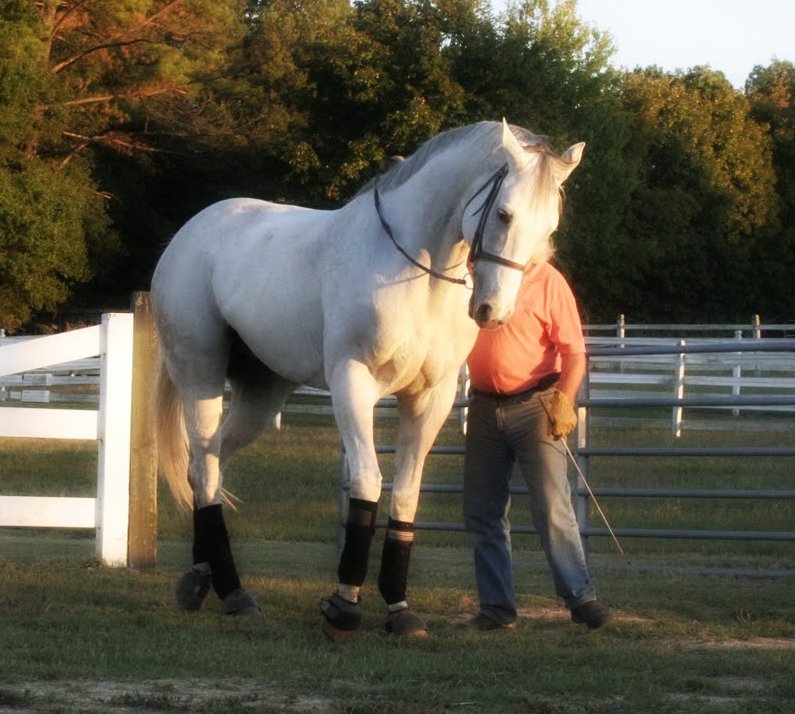
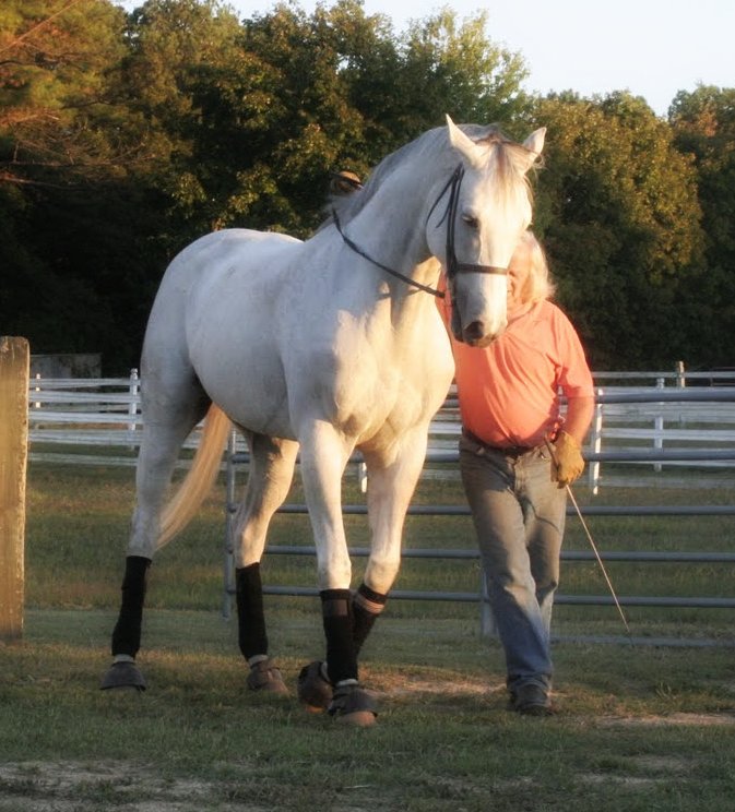
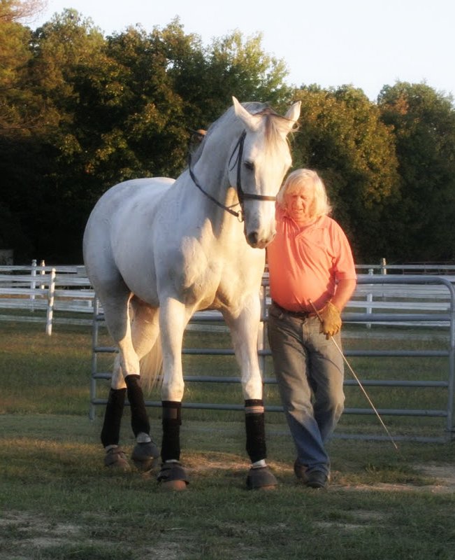
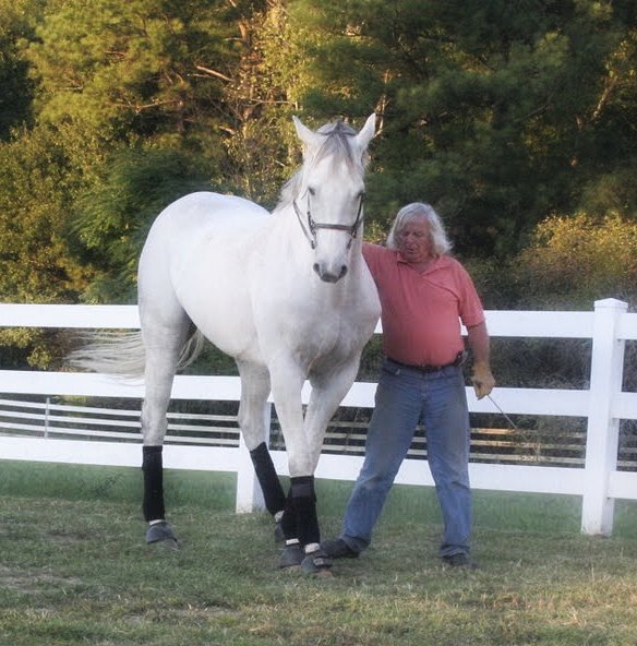
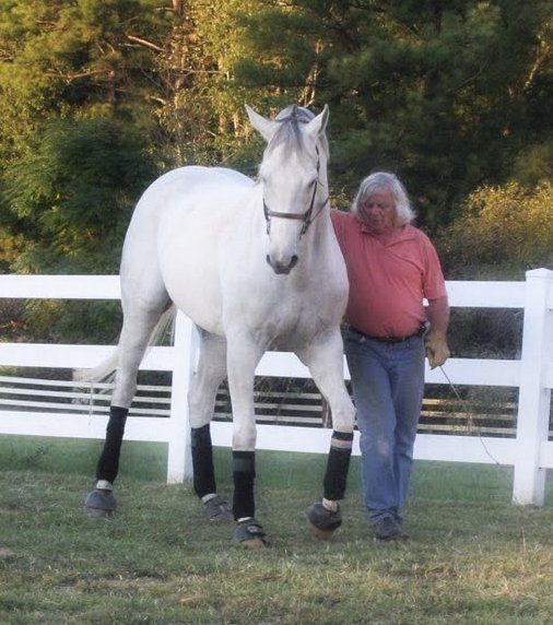
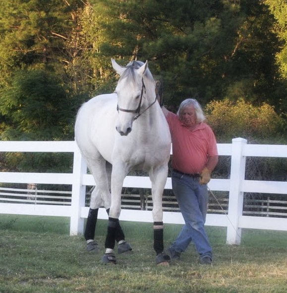
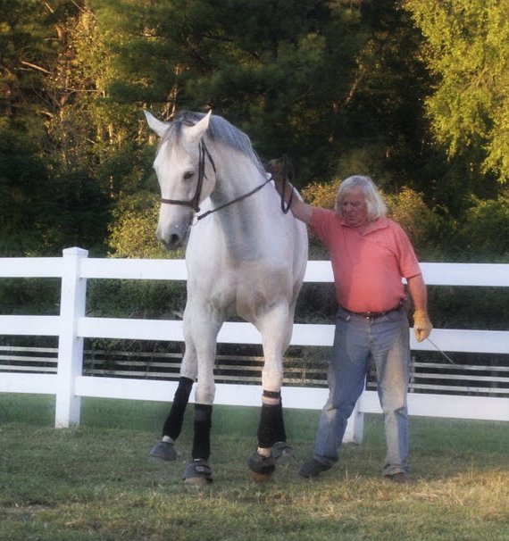
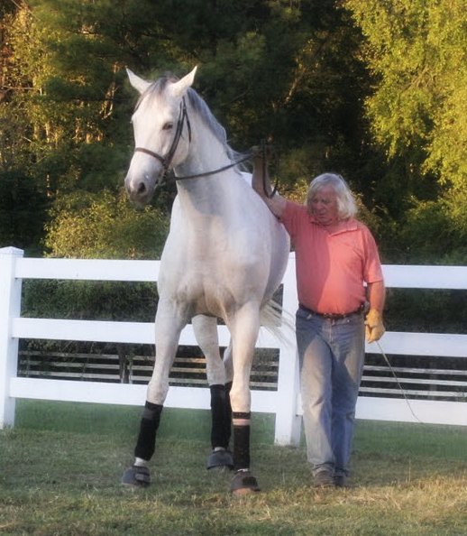
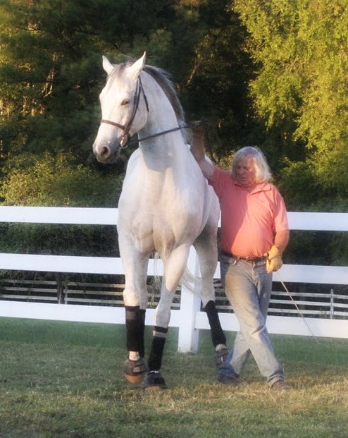
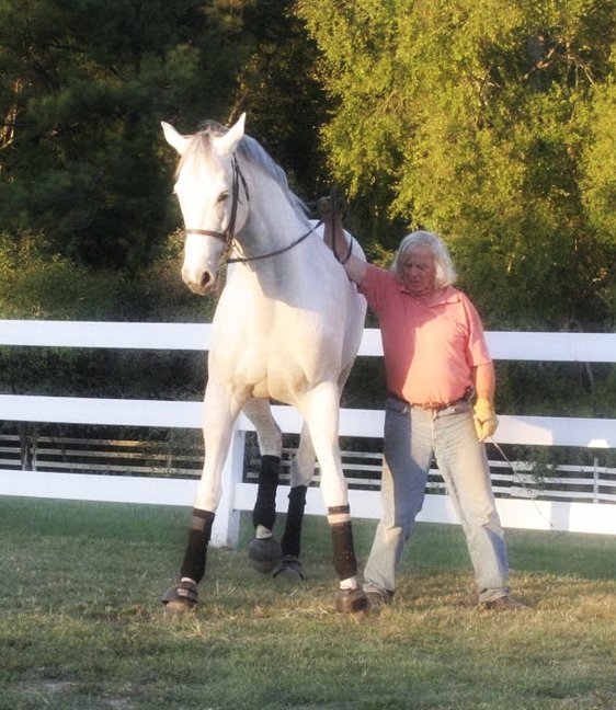
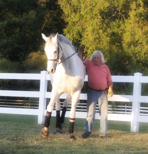
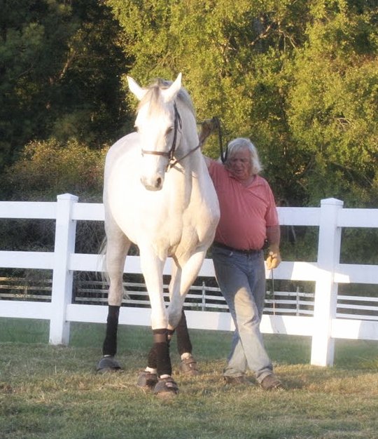
Helyn is delighted with Chazot's ongoing muscular development. She is taking picture of every muscle of his body and, Thanks to her artistic eye, the pictures are beautiful. Last night, during diner, Helyn asked me how you do it? My first thought was, I listen to him. My second thought was, I listen to him, so I responded, I listen to him. Helyn turned her right hand in circle, which is a gesture that Helyn does when she wants me to elaborate further. Well, I listen to him. Each day is different. As we walk around the ring for the warm up, Chazot's movements are telling me how the work needs to develop. For instance, yesterday we let Chazot play with the big ball. As a result, his shoulders were a little stiff today. I started the session focusing on suppleness. Chazot was quite receptive with the work. In fact his back felt quite good. I asked him to further control longitudinal flexion of his thoracolumbar spine and therefore balance control. We ended with a nice forward transmission of the force through the shoulders.
Proper muscular development is about harmony. A muscle never works alone. For each contraction there are compensatory contractions, muscles that stabilize the joints, others who redirect forces and so on. Vincent Van Gohg wrote, I dream my Painting and then I paint my dream. Each day the light influence the choice of the color, the intensity of the touch, etc. It is the same for the horse's education. Each day is different. Muscles develop influencing the horse's reaction. The choice of the move, the intensity or softness of the touch is not a preconceived opinion but instead a response the horse aiming at guiding the horse's brain toward the right coordination. Jean Luc Cornille www.scienceofmotion.com
Monday, January 23, 2012
Hind Legs Engagement and Stifle Problem
he thought that the horse's gait could actually be changed to rehab or prevent injuries is almost completely foreign to veterinarians as well as trainers.” (Betsy Uhl, DVM, PhD. 2011)
When equine locomotion and athletic performances are analyzed in great details, like under the microscope, limb kinematics abnormalities causing injuries can actually be corrected. The horse’s physique can actually be optimally coordinated for the athletic demand of the performance. This evolution is made possible by updated understanding of the horse’s physiology.
Born with advanced research studies, the Motion Microscope Therapy has mature into a different field of research; the training ring. The result is a powerful therapy identifying and addressing the source of the kinematics abnormality causing the injury.
“A major cause of lameness is lameness.” Rooney’s famous idea is that when it is repeated every stride a mild kinematics abnormality causes injury. Kinematics abnormalities may originate from morphological flaw or muscle imbalance, but also from training misconceptions. Lacking the support of adequate scientific knowledge great authors’ thoughts have been distorted over time and simplified to the point of meaningless formulas. In fact simplistic formulas are the main cause of equine injuries. In this series, we review, one by one, the kinematics abnormalities causing injuries and how training misconceptions can create such abnormalities.
Hind Legs’ Engagement.
Whatever the horse’s specialty, the base of all equine athletic performance is the engagement of the hind legs. The point here is not to question the need for hind legs’ engagement but instead to underline the fact that focusing on the hoof placement is a simplification, which places the horse at risk of injury. Sound locomotion demands precise coordination between forward swing of the hind limb around the hip joint and dorso-ventral rotation of the pelvis. In his quantitative study on Swedish Warmbloods comparing back and limbs kinematics of good and bad movers, Mikael Holmström observed greater pelvis rotation on above average movers. “The undulation of the pelvis was larger in the horses with good trot and increased in passage.” (1)
The hind limbs and the pelvis have to move in the same direction. When the hind limb swings forward, the pelvis rotates dorso-ventrally. When the hind leg moves backward into the pushing phase, the pelvis returns into a more horizontal position. Pelvis and limb movements are proportional but soundness demands their precise synchronization. The problem is that it is possible through whip or spurs to create deeper engagement of the hind leg without adequate pelvis rotation. The kinematics abnormality might please uneducated eyes but places the horse at risk of sacroiliac (SI) strain and stifle problem.
There is actually a strong recurrence of SI problems. One reason might be greater concern from the veterinary world for back problems.“Even if back soreness is thought to be only a compensation for hock pain and other musculoskeletal disorders, practitioners still have an obligation to evaluate and manage the back problem concurrently.” (2) The second reason might very well be the fast forward misconception that is currently rewarded in the show ring.
Some of the kinematics abnormalities leading to sacroiliac strain are similar to the kinematics abnormalities inducing stifle problems. The kinematics of SI injuries will be studied in detail during our next Immersion Program, (February 17, 18 & 19). In this discussion, we focus essentially on the misconceptions about hind leg engagement that is placing the horse at risk of stifle injuries.
If forced to do so, a horse not using the vertebral column properly will deeply engage the hind leg underneath himself furthering the forward rotation of the femur around the hip joint. While rotating around the hip joint, the femur undergoes simultaneously an inward rotation around the tibia. This rotary movement of the femur occurs toward the outside, (medial-to-lateral,) during the swing phase and toward the inside, (lateral-to-medial,) during the support phase.
As the protracting hind leg swings forward, the stifle extends and the usual medial-to-lateral rotating movement occurs. “If the extension is carried on beyond about 143-145°, there is a final lateral-to-medial twist, which rotates the patella medially and hooks the medial patellar ligament over the medial ridge of the femoral trochlea. The stifle is “locked” and flexion prevented.”(3) This is the mechanism of accidental locking of the patella. To unlock the stifle, the quadriceps muscle contracts, lifting the patella as the biceps contracts, pulling the patella laterally. The horse’s quick reflex contractions prevent accidental locking of the patella but induce stride after stride of abnormal stresses on the joint.
At the canter, the problem is unlikely to occur because the gait does induce longitudinal flexion of the horse’s thoracolumbar spine and the pelvis does oscillate dorso-ventrally. Both hind legs are moving together into the swing phase and the axis of rotation is the lumbo-sacral junction. At the contrary, at the trot as well as at the walk, one hind limb moves forward and the other moves backward. Each limb rotates around the hip joint and the pelvis “ducktail.” The dorso-ventral rotation is naturally reduced. When training misconceptions work toward stiffening the horse’s thoracolumbar column, dorso-ventral rotations of the pelvis are reduced even more and the horse increases the rotation of the femur around the hip joint.
On a video recorded along the dressage ring of the Atlanta Olympics for kinematics studies, several horses exhibited grotesque parodies during the medium walk. The horses over tracked the hind hooves, without adequate dorso-ventral rotation of the pelvis. They were moving at the walk like Tennessee walkers. The ducktail motion of the pelvis was accentuated and they further rotated the femur around the hip joint extending the stifle joint too far. Later during the dressage test, these horses demonstrated severe stifle pain during piaff and passage. Through manipulation, one can easily create dorso-ventral rotation of the pelvis.
The relation between pelvis rotation and overall flexion of the horse’s thoracolumbar spine is then apparent.Riding techniques altering longitudinal flexion of the horse’s thoracolumbar spine, therefore expose the horse to stifle injury. The most common misconceptions that stiffen the horse’s thoracolumbar column are speed and weight on the bit. A horse increases the speed by stiffening the thoracolumbar spine. The same reflex contraction is used by a horse leaning heavily on the bit. Lowering of the neck also tends to stiffen the back. Some horses have been shown to lose vertebral mobility in the thoracic area and all horses are losing vertebral mobility in the lumbar area when the neck is lowered.
The horse’s adaptation to the rider’s weight is to increase the duration of the hind limb’s supporting phase. More exactly, the horse increases the duration of the decelerating phase. This of course demands a more forward placement of the hind leg at impact. However, efficiency does not relate to the hoof placement but instead to how well each joint of the alighting hind limb is placed to optimally absorb impact forces. At the piaff for instance, the horse places the alighting hind leg less forward under the body than during collected trot. Uneducated riders think otherwise but their opinions lack understanding of the performance’s athletic demand.
While the hind and front limbs are acting like a lever at the walk, they work more like a spring at the trot. The vertical position of the hind limb under the croup is best suited for the task of decelerating the horse’s body through the flexion of the joints. “The hind legs have a considerable braking activity to avoid forward movement of the body over the forelegs. The forelimbs have a larger propulsive activity.”(4) As the joints of the supporting hind leg fold, they resist gravity and forward displacement of the body over the forelegs. Gravity and inertia forces are loading the supporting hind leg. If the hoof was placed more forward under the body, the stress on the canon bone would be greater. Also, the hock’s middle joint T3-TC would be under excessive stress and therefore prone to arthritis.
The opposite is equally damaging. When the supporting hind leg alights behind the vertical, the stress is greater on the hock’s lower joint, Mt3-T3. Basically the horse is placed in the situation of functional straight hock and is prone to injuries related to the morphological flaw. Horses are kind enough to perform even if the rider places their body into stressful positions. The price of course is lameness. More and more research studies demonstrate that cartilage issues, such as arthritis in the hock, result from abnormal stresses on tarsal bones. In most instances you see bone damage before you see the cartilage changes.
The horse adapts the hind legs’ hoof placement to the athletic demand of the performances. During the stride preceding the flying change, the horses achieving the best performances increase the length of time that both hind hooves remain on support. Basically, they increase the decelerating phase of the hind legs. “Preceding a lead change, the higher-scoring horses increased their contact duration of the hind limbs and decreased the length of step and time between forelimb impacts to prepare to execute the lead change in the succeeding airborne phase.” (5)
The race horse engages the hind legs more forward under his body than the dressage horse. Such engagement is definitively helped by the longitudinal flexion of the thoracolumbar spine and dorso-ventral rotation of the pelvis and sacrum around the lumbo-sacral junction, which is all natural at the canter. However, the race horse does not utilize greater engagement of the hind legs to enhance balance. Instead, the race horse utilizes the elastic strain energy accumulated during the decelerating phase to maximize the propulsive action.
The horse’s morphology also does influence the position of the hind hoof under the body at impact. For instance, two horses working with the same thoracolumbar column and pelvis rotation; a horse with a sickle hock will place the hind hoof more forward while a horse with a straight hock will place the hind hoof less forward. Forcing a straight-legged horse to track up deeply, would place the horse at risk of hock injury as well as sacroiliac problems and/or stifle issues.
Simplicity is the greatest achievement of knowledge but simplicity without knowledge is the greatest cause of equine injuries. “The horse’s hind legs need to track up at working trot”, is the type of simplistic formula which, if applied without sound understanding of the horse’s vertebral column mechanism and pelvis rotation, is likely to cause injury. In this circumstance the cure is knowledge. Greater engagement of the hind legs is not the cause but instead the result of sophisticated body coordination. This body coordination cannot be created by acting directly on the hind legs. Training formulas are, for a great part, grossly inaccurate and the rider’s knowledge of the underlying biomechanics factors is the horse’s best chance of soundness.
James Rooney identified the kinematics abnormalities causing injuries. The pathologist pioneered the biomechanics of lameness. The MotionMicroscope Therapy is about identifying and correcting the source of the kinematics abnormalities causing injuries. The Motion MicroscopeTherapy pioneers the biomechanics of soundness.
Jean Luc Cornille
Copyright 2012
References,
(1) (Mikael Holstrom, Quantitative study on conformation and trotting gaits in the Swedish Warmblood riding horse. Dissertation, Uppsala, 1994)
(2) (Kevin K. Haussler, DVM, DC, PhD, Preface, Veterinary Clinics of North America 1999)
(3) (James R. Rooney, Biomechanics of lameness in horses, 1976)
(4) (Erid Barrey, Sophie Biau, Locomotion of dressage horses, Conference on Equine Sport Medecine and Science, 2002)
(5) (N. R. Deuel, PhD: J. Park, PhD, Canter lead change kinematics of superior Olympic dressage horses, 1990)
http://www.scienceofmotion.com/motion__microscope_therapy_.html
Tuesday, January 17, 2012
Equine Back Research

History of Equine Back Research Studies
Since the inception of the Immersion Programs I have observed that going through the history of equine research studies has better helped riders, trainers and therapists to understand how the horse’s vertebral column effectively functions. This history affords the participants a greater accuracy as well as an evolution from the simplicity of past theories to the complexity of actual knowledge. Studying the past also reveals from where and when the theories, that are still promoted in these present days come from, and just how long it has been since they have progressed. This text is, of course, just a brief summary of the original lecture.
It is true that the fascicles of the main back muscles are inserted obliquely on the dorsal spines The fascicles of the longissimus dorsi muscles are oriented oblique, down and forward while the fascicles of the spinaleus dorsi and more exactly the multifidius, are oriented oblique, down and backward. Their action induces rotary forces on the dorsal spines and correspondent vertebrae. This was explained by E. J. Slijper in 1946. The Dutch scientist also described the horse’s vertebral column functioning as a “bow” that can be flexed by the action of the “string”, which is composed of pectoral and abdominal muscles. The theory was referred to as the “bow and string concept.” With some variables, this is basically the concept behind most actual riding techniques as well as the concept supported in the video that started the discussion. The problem is that the concept was presented in 1946. Scientific findings and therefore knowledge has greatly evolved since 1946.
In 1964 Richard Tucker explored the thought that acting on the dorsal spines, the back muscles were allowing the vertebrae to transmit the thrust generated by the hind legs into horizontal forces, (forward movement), and to create vertical forces resisting gravity and therefore permitting balance control. Tucker furthered Slijper’s description explaining how, through their insertion on the dorsal spines, the muscles were compressing the vertebrae against each other favoring forward transmission of horizontal forces, (forward movement.) Simultaneously back muscles are inducing rotary movements of the vertebrae creating vertical forces, (resistance to gravity and balance control.) Tucker moved away from the simplistic idea that the thoracoumbar column was flexing and extending as a whole. The Polish scientist pointed out that due to the curvature that characterizes the shape of the horse’s thoracolumbar spine, the vertebrae and muscles situated on the ascending side of the curvature were working in the opposite way than the vertebrae and muscles situated on the descending side of the thoracolumbar curvature.
In 1969, James Rooney demonstrated that the work of these muscle groups, which are arranged in mirror images, has to be perfectly synchronized to ensure proper locomotion. If the fascicles of the longissimus muscles contract first, the thoracic spine extends. If the fascicles of the spinaleus dorsi contract first, the lumbar spine stiffens. Rooney basically demonstrated the damage created by the shifts of the rider’s weight, which is a theory that is still emphasized in modern days. If the rider’s weight is acting back to front, as emphasized in the driving seat theory, the rider stiffens the horse’s thoracic vertebrae. By contrast, if the rider’s weight is acting front to back, the rider stiffens the horse’s lumbar vertebrae. Rooney’s findings suggested an equitation based on a rider’s body maintained constantly on a neutral balance, which is exactly vertical over the seat bones. One of the defenders of the long and low theory referred to “Pilates.” If this person really knew Joseph Pilates’ approach, she would have realized that maintaining the rider’s body in perfect neutral balance and therefore with the vertebral column almost straight is Pilates’ real teaching. Her perception of Pilates for the horse is that the abdominal muscles flex the thoracolumbar spine. This is not Pilates’ teaching. The real Pilates idea is to balance the work of both abdominal and back muscles to straighten the spine.
Rooney’s work also suggested that the real relation between the horse’s vertebral column and the rider’s back was more at the level of subtle movements of the rider’s back instead of shifts of the rider’s weight. In relation to the work of the back muscles, Rooney’s explanation differed from Tucker’s view. However, understanding how the horse’s vertebral column converts the thrust generated by the hind legs, which is basically a horizontal force, into forces resisting gravity, which are vertical forces, is easier to visualize mentally with Tucker’s explanation. This does not mean that Tucker’s explanation should be taken word for word. All these explanations are attempting to describe forces, which is an abstract concept. True understanding demands several ideas aiming toward the same concept. Rooney’s insight was that the creation of upward vertical forces through the spine was achieved by the direction of the muscles’ work without inducing much movement of the vertebrae.
The pathologist explained that in order to create horizontal forces, (forward movement,) and vertical forces, (resistance to gravity and balance control), two muscles are needed, one acting horizontally and one acting vertically, or, a single muscle acting in an oblique manner. Such insertion allows the same muscle to create both horizontal and vertical forces. This is exactly how the fascicles of the main back muscles are oriented and function. This was the beginning of a long series of research aiming toward a functioning of the horse’s back muscles based on the subtle management of forces instead of increasing the movements of the vertebrae. This was 1969 and we were, at that time, already far away from the infantile idea that a single action such as lowering the neck could flex the whole thoracolumbar spine and also that gaits and performances can be improved by increasing the range of motion of the horse’s thoracolumbar column.
Rooney also questioned the veracity of the bow and string concept. As a pathologist, Rooney observed firsthand the discrepancy between the large mass and power of the back muscles and small mass and limited power of the abdominal muscles. We have recently published a picture showing a cut-away of the back muscles and by comparison a cut-away of the abdominal muscles executed at the same vertical plain. Rooney basically demonstrated that abdominal muscles do not have the capacity to flex the back muscles. Longitudinal flexion of the horse’s thoracolumbar spine is instead, created by the precise coordination of the main back muscles that are situated above the vertebral bodies.
Rooney’s work suggested that the contraction of abdominal muscles would assist the flexion of the back but not create it. Instead of lowering the horse’s neck and stimulating hind legs engagement, as suggested in the video, the flexion of the horse’s thoracolumbar column is more likely to occur through harmonic motion of the rider’s back influencing the work of the horse’s back muscles. Three and half decades later, uneducated trainers and riders are promoting the lowering of the neck as a new way to engage the horse’s back.
Even if equine research studies were brought to a complete halt, the practical application of available knowledge would considerably enhance the horse’s performances and in particular the horse’s soundness. Very few of today’s advanced scientific discoveries benefit the horse through better training and riding techniques. The reason is that instead of questioning old ideas in the light of new findings trainers, riders and judges are integrating new discoveries to old beliefs. Considering the cost of raising, maintaining and training a horse, it is incomprehensible that the practical application of modern science, which could greatly prolong and further the horse’s career, preserve the horse’s soundness and consequently cut the vet bills, is rejected in favor of archaic but familiar approaches.
In1976 was also when Hans Carlson demonstrated that the main function of the back muscles was not to increase the range of movement of the horse’s vertebral column, as suggested in the video as well as in the show ring, but at the contrary, to protect the vertebral column from movements exceeding the thoracolumbar spine’s possible range of motion. Uneducated riders argue that the study was made on cats. Carlson’s study was effectively effectuated on cats, which demonstrates in fact, that visual impressions can easily lead to the wrong perception. Multiple studies have then been done duplicating the same protocol and the findings were similar with horses and most terrestrial mammals. Basically, all the theories promoting better performances and gaits through stretching and greater amplitude of the horse’s vertebral column movements are in direct contradiction with the way the horse’s vertebral column and surrounding muscles are designed to work.
In 1980, Leo Jeffcott measured the range of possible movement of the horse’s vertebral column. Many studies after Jeffcott found differences in the location of vertebral column movements but they all found a limited range of motion. Basically, the back muscles do not increase the vertebral column range of movement but, at the contrary, resist forces induced on the horse’s vertebral column in order to maintain the vertebral column movements within the limits of its possible range of motion. This was 1980 and it was already demonstrated that theories such as the swinging back and stretching were in plain contradiction with the way the horse’s vertebral column and back muscles operate. At this point of knowledge, the thought that the horse’s thoracolumbar column was flexing longitudinally and laterally as a whole was totally blown away. All investigations clearly demonstrated that while greater movements were possible between T9 and mostly T14, while some horses show mobility until T16, the rest of the horse thoracolumbar spine was quite rigid. Movements occur but within the limits of a restricted ranged of motion.
In 1999, Jean Marie Denoix published a comprehensive study on the functioning of the horse’s vertebral column. Among the very pertinent discoveries was the way each vertebra rotates in relation to the other. Denoix’s research presented a work of the back muscles quite different than previously believed. Tucker for instance, theorized that lateral bending induced pressure on the inside side of the vertebrae. Denoix demonstrated that in fact lateral bending was created by a rotation of one vertebrae around the other. The French author was the first to present a comprehensive study about the fact that lateral bending is always coupled with a movement of transversal rotation and that rotation is, also, always associated with lateral bending.
In line with James Rooney, Denoix’s work demonstrated that gaits and performances were the outcome of back muscles creating and orchestrating forces instead of inducing greater range of motion of the vertebrae. Before the birth of the 21st century, scientific research had already demonstrated that all forms of equitation based on increasing the horse’s vertebral column range of motion was not only antiquated but contrary to the way the horse’s vertebral column effectively works.
2003 commenced better understanding of the plasticity of muscles’ function. Muscles’ architecture is now understood at a much deeper level. The involvement of the horse’s vertebral column into gaits and performances is more about muscles creating and orchestrating forces than muscles moving vertebrae. For centuries, horses have been forced to execute movements of the back that their vertebral column was not designed to create. In fact, they did not increase the range of motion of their vertebral column for the very simple reason that their vertebral column mechanism does not allow greater movement. Instead, they found ways to compensate for the incongruity of their riders’ demand. The best horses did not increase the amplitude of their vertebral column’s movement but instead, the subtle coordination of forces, giving to the rider an impression of ease that the rider interpreted as relaxation, stretching, swinging back and other misconceptions. Some horses have succeeded to figure, within the incoherency of the rider’s stimulus, how to orchestrate more or less appropriately their physique for the athletic demand of the performance. Many others have tried as hard as their talented peers but nature did not give them the same athletic and mental abilities and they succumbed to lameness.
One of the most common deceptions is the belief that the lowering of the neck flexes the lumbar vertebrae and increases their range of motion. The optical illusion was explained in 1986 by Jean Marie Denoix. The lowering of the neck reduces the mobility of the lumbar vertebrae. This is true for every horse. Stiffening of the lumbar vertebrae hampers proper dorso-ventral rotation of the pelvis and therefore sound kinematics of the hind legs. In order to compensate for the stiffening of the lumbar vertebrae, the horse increases the work of the iliopsoas muscles, which swings the hind limbs forward. Since the iliopsoas is placed under the lumbosacral junction, increased work of the iliopsoas muscle does induce greater rotation of the lumbosacral junction. This lumbosacral rotation does give the optical illusion that the whole lumbar region moves. In fact, the lumbar vertebrae do not flex. Instead, the horse compensates for the rigidity of the lumbar spine, that was created by the lowering of the neck, with greater intensity in the lumbosacral junction that is situated behind the lumbar vertebrae. The theories of relaxation, stretching and greater mobility of the vertebral column are naïve interpretations of a mechanism which in fact, is working exactly the opposite way.
Today’s knowledge allows returning the favor to the horse. The practical application of advanced scientific knowledge permits us to understand how the horse’s back effectively functions and how the rider can guide the horse’s brain toward efficient coordination of the vertebral column mechanism. This is done by reducing the range of motion of the rider’s back and matching the range of motion of the horse’s back. Through a subtle body language, the rider guides the horse’s brain toward the body coordination appropriate for the effort. This is classical training. At the 17th century The Duke of Newcastle already talked about the stability of the rider’s pelvis. The classic author uses the terms “unmovable pelvis.”
The practical application of pertinent scientific discoveries commences by questioning old theories in the light of new knowledge. It is astounding that instead of upgrading their training and riding techniques to the findings of modern research studies, trainers deliberately refuse new knowledge under the name of tradition. They submit their horses to the same incongruities and suffering as the horses of previous generations. It is equally astounding that riders follow and even protect these primitive ideas. Fortunately, there are also trainers who evolve, upgrading their techniques with true knowledge. In the same line of thought, there are riders who do not let their horses to be damaged by submissive and uneducated training techniques. One of the great horses that we have in training right now had the luck to belong to a rider who refused poor training techniques. The rider stood up for her horse and the horse, which was born as a good horse has evolved into a great horse.
We have today, the capacity to prepare efficiently the horse’s physique for the athletic demand of the performance. We owe this knowledge to the horse. Refusing progress and perpetuating old and heretic beliefs is not classic; it is archaic. If one wants to protect the horse from rollkur, draw reins, deep work and other exploitations, one needs to evolve from the common denominator of all these poor training techniques, which is ignorance. A real classic trainer learns how the horse’s body effectively works and then applies the motto of the most classical school of riding. “Respect for tradition should not prevent the love of progress.” (Colonel Danloux, Cadre Noir de Saumur, 1931)
Jean Luc Cornille
References:
- E. J. SLijper. Comparative biologic-anayomical investigations on the vertebral column and spinal muscles. Verhandelingen der kononklijke Nederlandsche Akademia van Wetenschappen, AFD, Natuurkunde. Tweede Sectie, deel XLII, #5
- Richard Tucker, Contribution to the Biomechanics of the vertebral column. Rotary system induced in the thoraco-lumbar curvature by the epacial musculature. Acta Theriologica, Vol. IX, 12: 171-193. Bialowieza. 30. XI. 1964
- James R. Rooney DVM, Biomechanics of lameness in horses. The William and Wilkins company, Baltimore, 1969
- Hans Carlson, Halbertsma J. and Zomlefer, M. 1979, Control of the trunk during walking in the cat. Acta physiol. Scand. 105, 251-253
- Leo B. Jeffcott, Natural Rigidity of the horse’s backbone, 1980. Equine vet J. 1980, 12 (3) , 101-108
- Jean Marie Denoix, DVM. PhD, Spinal Biomechanics and Functional Anatomy, 1999
Thursday, December 01, 2011
Educate your Eye
Shoulder in executed in hand with correct and inverted rotation.
Even if the concept of rotation associated with lateral bending has been clearly explained in 1999 with Jean Marie Denoix’s magisterial work, the concept remains foreign to judging standards and most training techniques. The concept is important to know; proper or inverted rotation is often the difference between good and poor performance as well as soundness or limbs injuries and back issues. In practically all the rehabilitations that we have completed, the subject of inverted rotation was part of the problem.
The first picture series shows right shoulder in and then right turn, executed in hand with correct rotation. The direction of the rotation is illustrated by a white arrow. At the second frame, Chazot is putting some weight on the bit while lowering the neck. The gesture increases the load on the forelegs. Chazot is then almost associating right lateral bending and inverted rotation. Fortunately, His mind was on the correct coordination and the fault lasts only one frame. He corrects himself at the next frame.
Correct Shoulder In
Right laterial bending coupled with correct rotation


Incorrect Shoulder In
Right laterial bending coupled with inverted rotation


The second picture series shows right shoulder in executed in hand and coupled with inverted rotation. The direction of the rotation is illustrated with a red arrow. The good thing with chazot is that he never does anything half way. He is trying to put some weight on the bit and the rotation become worse each step. Right shoulder in and therfore right laterial bending coupled with inverted rotation clearly shift the weight on the horse's left shoulder.
In fact the series presented first as the good rotation is the continuation of this bad series. Chazot recovered his balance and was able to come back with the correct coordination between lateral bending and proper rotation.
One may ask how inverted rotation does feel. The feeling is easy to recreate. One just has to halt the horse on the rail keeping the horse’ vertebral column straight and bending the horse neck laterally toward the inside of the ring, at an angle of about 90°. As the horse bends the neck, the rider will be shifted toward the outside of the horse’s back and on the outside seat bone. This is the feeling of inverted rotation.
Jean Luc Cornille Copyright©2011
Subscribe to:
Comments (Atom)